Gross shows cut-opened specimen of the right kidney showing a unilocular hydatid cyst with multiple daughter cysts.
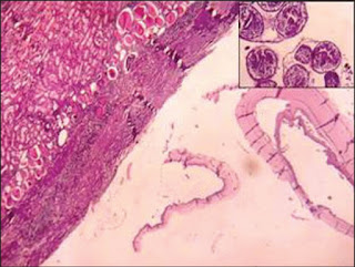
Microscopy shows laminated membranes of hydatid cyst with adjacent renal tissue showing compression and features of chronic pyelonephritis. (H and E, 100×). (Inset) Brood capsules showing scolices and lanceolate hooklets (H and E, 450×).
Impression: Isolated renal hydatid cyst.
Note: For further reading refer Kamath AS, Rao SP et al Isolated Renal Hydatid Cyst, Journal of Dr. NTR University of Health Sciences 2014;3(1): 70-71


No comments:
Post a Comment